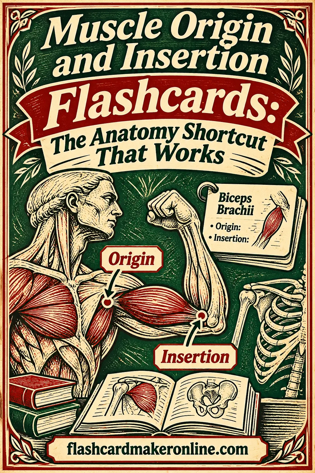
Muscle Origin and Insertion Flashcards for PT and Massage School
The fastest way to learn muscle origins and insertions is one card per muscle: the muscle name on the front, and its origin, insertion, action and nerve on the back, always in the same order. Group those cards by region, study one region at a time, and add a few reverse cards that start from an action ("Which muscles flex the elbow?") so you can answer questions from either direction.
You can build that whole deck in a free flashcard maker with no account, study it on screen, and print it double-sided. Below you'll find the card layout that works, a 65-muscle sample deck you can paste in, memory tricks, and a six-week schedule.
One note before you start: textbooks don't all describe attachments the same way. One book says serratus anterior starts on ribs 1 to 8, another says 1 to 9. Use the wording from your own course textbook and your instructor's lists, since your exam is graded against them.
Why Origins and Insertions Are So Hard to Memorize
Muscle attachments feel like a wall of random words at first. Here's why, and what helps.
The volume is huge. A kinesiology or gross anatomy course can ask for dozens of muscles, each with three to five facts, and many of them sound alike.
The bony landmarks repeat. The greater tubercle, the linea aspera and the ischial tuberosity show up on card after card, so they blur together.
Lists don't show the logic. When you read a table of origins and insertions top to bottom, every row looks equally arbitrary. In reality, attachments follow patterns. Muscles that share an origin often share an action. A muscle's action follows from where it crosses a joint and which way it pulls.
Recognition feels like knowing. Rereading notes feels productive because the words look familiar, but your exam asks you to produce the answer from memory. Flashcards close that gap by making you recall before you check. The active recall method explains why that effort is what makes facts stick.
Quick refresher: origin, insertion, action
Before you write a single card, get the three terms clear. Most courses teach them like this:
| Term | Usual definition | Quick way to think about it |
|---|---|---|
| Origin | The attachment that usually stays more fixed during movement, often the more proximal one | The anchor |
| Insertion | The attachment that usually moves more, often the more distal one | The part that gets pulled |
| Action | The movement the muscle produces when it contracts | What happens when the insertion is pulled toward the origin |
| Innervation | The nerve that supplies the muscle | Who sends the signal |
Once you see an action as "insertion pulled toward origin," you can often figure it out instead of memorizing it. Biceps brachii starts at the scapula, crosses the front of the elbow and inserts on the radius. Pull the radius toward the shoulder and the elbow bends. With the palm down, the tendon wraps around the radius, so pulling on it spins the forearm palm up. Flexion and supination aren't two random facts; they're what that line of pull has to do.
The Card Layout That Works
The single biggest decision is how much to put on each card. There are three good layouts, and most students end up using all three at different stages.
| Layout | Front | Back | Best for |
|---|---|---|---|
| Full muscle card | Muscle name | Origin, insertion, action, nerve, in a fixed order | Your main deck; learning each muscle as a whole |
| Split cards | "Supraspinatus, insertion?" | Greater tubercle, superior facet | Muscles you keep missing; breaking a hard card into pieces |
| Reverse (action) cards | "Muscles that laterally rotate the humerus" | Infraspinatus, teres minor, posterior deltoid | Exam questions that start from a movement or a landmark |
Layout 1: the full muscle card
This is the core of the deck. Put just the muscle name on the front. On the back, list four short lines in the same order every time:
- O (origin)
- I (insertion)
- A (action)
- N (nerve)
A fixed order matters. When every back follows O, I, A, N, you can recite in that order and notice exactly which piece you forgot. If your course doesn't test innervation, drop the N line.
Here's what a full card looks like:
Front: Infraspinatus Back: O - infraspinous fossa of scapula. I - greater tubercle of humerus (middle facet). A - laterally rotates arm; helps stabilize the shoulder. N - suprascapular nerve.
When you review, say the whole back out loud before you flip. Only count the card as correct if you got every line. Partial credit is how weak cards hide.
Layout 2: split cards for trouble spots
Some muscles have long, messy attachments. The pectoralis major origin, the latissimus dorsi origin and the adductor magnus insertion are classic examples. If you keep missing one piece of a full card, don't just keep flipping it. Split that muscle into two or three smaller cards:
Front: Latissimus dorsi, origin? Back: Spinous processes of T7 to T12, thoracolumbar fascia, iliac crest, lower 3 or 4 ribs.
Smaller cards are easier to get fully right, which is the point. Once you can answer the split cards cold, go back to the full card. The minimum information principle is the reasoning behind this: a card should ask one thing whenever it can.
Layout 3: reverse cards for movements and landmarks
Exams and clinical work often ask the question backwards: a practical points to a landmark, or a client says it hurts to lift their arm out to the side. Reverse cards train this direction:
Front: What attaches to the ischial tuberosity? Back: Biceps femoris (long head), semitendinosus, semimembranosus, hamstring part of adductor magnus.
Front: Prime movers of hip abduction Back: Gluteus medius and gluteus minimus (helped by tensor fasciae latae).
You don't need a reverse card for every muscle. Make them for the landmarks and movement groups your instructor emphasizes.
Abbreviations to keep backs short
A few standard abbreviations keep backs short. Use them the same way on every card:
| Abbreviation | Meaning |
|---|---|
| ASIS / AIIS | Anterior superior / anterior inferior iliac spine |
| PSIS | Posterior superior iliac spine |
| SP / TP | Spinous process / transverse process |
| MC / MT | Metacarpal / metatarsal |
| IT band | Iliotibial tract |
| CN XI | Cranial nerve 11 (spinal accessory) |
Sample Deck: Shoulder and Upper Limb
Here's a ready-made upper limb section. Each back follows the O, I, A, N order. The fronts are just the muscle names, so you can paste them straight into the maker's "Paste a list" box using the front : back format shown later. Attachments are written the way many standard anatomy texts describe them; check them against your course book and adjust the wording to match.
| Front | Back |
|---|---|
| Trapezius | O - external occipital protuberance, superior nuchal line, ligamentum nuchae, SP of C7 to T12. I - lateral third of clavicle, acromion, spine of scapula. A - upper fibers raise (elevation) and upwardly rotate scapula; middle retract; lower depress. N - CN XI. |
| Levator scapulae | O - TP of C1 to C4. I - medial border of scapula from superior angle to root of spine. A - raises scapula (elevation), downwardly rotates it. N - dorsal scapular nerve and cervical nerves C3 to C4. |
| Rhomboid major | O - SP of T2 to T5. I - medial border of scapula from root of spine to inferior angle. A - retracts and raises scapula, downwardly rotates it. N - dorsal scapular. |
| Serratus anterior | O - outer surfaces of upper 8 or 9 ribs. I - anterior surface of medial border of scapula. A - protracts and upwardly rotates scapula, holds it against the chest wall. N - long thoracic. |
| Pectoralis minor | O - ribs 3 to 5 near the costal cartilages. I - coracoid process. A - depresses and protracts scapula, stabilizes it. N - medial pectoral. |
| Pectoralis major | O - medial half of clavicle; sternum and costal cartilages 1 to 6; external oblique aponeurosis. I - lateral lip of intertubercular sulcus. A - adducts and medially rotates arm; clavicular head flexes. N - lateral and medial pectoral. |
| Latissimus dorsi | O - SP of T7 to T12, thoracolumbar fascia, iliac crest, lower 3 or 4 ribs. I - floor of intertubercular sulcus. A - extends, adducts and medially rotates arm. N - thoracodorsal. |
| Deltoid | O - lateral third of clavicle, acromion, spine of scapula. I - deltoid tuberosity of humerus. A - abducts arm; anterior fibers flex and medially rotate; posterior fibers extend and laterally rotate. N - axillary. |
| Supraspinatus | O - supraspinous fossa. I - greater tubercle, superior facet. A - starts abduction of arm, stabilizes shoulder. N - suprascapular. |
| Infraspinatus | O - infraspinous fossa. I - greater tubercle, middle facet. A - laterally rotates arm. N - suprascapular. |
| Teres minor | O - upper two-thirds of lateral border of scapula. I - greater tubercle, inferior facet. A - laterally rotates arm. N - axillary. |
| Subscapularis | O - subscapular fossa. I - lesser tubercle. A - medially rotates arm. N - upper and lower subscapular. |
| Teres major | O - dorsal surface of inferior angle of scapula. I - medial lip of intertubercular sulcus. A - medially rotates, adducts and extends arm. N - lower subscapular. |
| Coracobrachialis | O - coracoid process. I - medial shaft of humerus, middle third. A - flexes and adducts arm. N - musculocutaneous. |
| Biceps brachii | O - short head from coracoid process; long head from supraglenoid tubercle. I - radial tuberosity and bicipital aponeurosis. A - flexes elbow, supinates forearm, assists shoulder flexion. N - musculocutaneous. |
| Brachialis | O - distal half of anterior humerus. I - coronoid process and tuberosity of ulna. A - flexes elbow in any forearm position. N - musculocutaneous (small part from radial). |
| Triceps brachii | O - long head from infraglenoid tubercle; lateral and medial heads from posterior humerus. I - olecranon. A - extends elbow; long head helps extend and adduct arm. N - radial. |
| Brachioradialis | O - upper two-thirds of lateral supracondylar ridge of humerus. I - lateral distal radius just above styloid process. A - flexes elbow, strongest with forearm in neutral. N - radial. |
| Pronator teres | O - medial epicondyle and coronoid process of ulna. I - middle of lateral surface of radius. A - pronates forearm, assists elbow flexion. N - median. |
| Supinator | O - lateral epicondyle, radial collateral and annular ligaments, supinator crest of ulna. I - proximal third of radius. A - supinates forearm. N - deep branch of radial. |
| Flexor carpi radialis | O - medial epicondyle. I - base of 2nd MC (and 3rd). A - flexes wrist, radially deviates it. N - median. |
| Flexor carpi ulnaris | O - medial epicondyle; olecranon and posterior ulna. I - pisiform, hook of hamate, base of 5th MC. A - flexes wrist, ulnar deviates it. N - ulnar. |
| Flexor digitorum superficialis | O - medial epicondyle, coronoid process, anterior radius. I - sides of middle phalanges of fingers 2 to 5. A - flexes PIP joints, helps flex MCP joints and wrist. N - median. |
| Flexor digitorum profundus | O - anterior and medial ulna, interosseous membrane. I - bases of distal phalanges of fingers 2 to 5. A - flexes DIP joints, helps flex the rest of the finger and wrist. N - ulnar (medial part) and anterior interosseous branch of median (lateral part). |
| Extensor carpi radialis longus | O - lateral supracondylar ridge. I - dorsal base of 2nd MC. A - extends wrist, radially deviates it. N - radial. |
| Extensor digitorum | O - lateral epicondyle. I - extensor expansions of fingers 2 to 5. A - extends fingers, assists wrist extension. N - posterior interosseous. |
| Extensor carpi ulnaris | O - lateral epicondyle and posterior ulna. I - base of 5th MC. A - extends wrist, ulnar deviates it. N - posterior interosseous. |
That's 27 upper limb cards. Notice the patterns hiding in the table. Most wrist and finger flexors start on or near the medial epicondyle, the common flexor origin. Most wrist and finger extensors start on or near the lateral epicondyle, the common extensor origin. Three of the four rotator cuff muscles land on the greater tubercle, and the one that doesn't, subscapularis, is the one that rotates the arm medially. Spotting these groups turns 27 facts into a handful of rules plus exceptions.
Sample Deck: Hip and Lower Limb
The lower limb has fewer muscles than the upper limb in most course lists, but the attachments are bigger and the landmarks get mixed up easily. Pay special attention to the pelvis: ASIS, AIIS, the ischial tuberosity and the pubic rami each anchor a different muscle group.
| Front | Back |
|---|---|
| Psoas major | O - bodies and TP of T12 to L5 and the discs between them. I - lesser trochanter. A - flexes hip; helps flex the trunk. N - anterior rami of L1 to L3. |
| Iliacus | O - iliac fossa. I - lesser trochanter (with psoas tendon). A - flexes hip. N - femoral. |
| Gluteus maximus | O - posterior ilium, dorsal sacrum and coccyx, sacrotuberous ligament. I - IT band and gluteal tuberosity of femur. A - extends and laterally rotates hip. N - inferior gluteal. |
| Gluteus medius | O - outer surface of ilium between anterior and posterior gluteal lines. I - lateral surface of greater trochanter. A - abducts and medially rotates hip; keeps pelvis level in single-leg stance. N - superior gluteal. |
| Gluteus minimus | O - outer surface of ilium between anterior and inferior gluteal lines. I - anterior surface of greater trochanter. A - abducts and medially rotates hip. N - superior gluteal. |
| Tensor fasciae latae | O - ASIS and anterior iliac crest. I - IT band, which continues to the lateral tibial condyle. A - flexes, abducts and medially rotates hip; tenses IT band. N - superior gluteal. |
| Piriformis | O - anterior surface of sacrum. I - superior border of greater trochanter. A - laterally rotates the extended hip; abducts the flexed hip. N - nerve to piriformis (S1 to S2). |
| Sartorius | O - ASIS. I - superior medial tibia (pes anserinus). A - flexes, abducts and laterally rotates hip; flexes knee. N - femoral. |
| Rectus femoris | O - AIIS and ilium just above the acetabulum. I - patella, then tibial tuberosity via patellar ligament. A - extends knee, flexes hip. N - femoral. |
| Vastus lateralis | O - greater trochanter and lateral lip of linea aspera. I - patella, then tibial tuberosity. A - extends knee. N - femoral. |
| Vastus medialis | O - intertrochanteric line and medial lip of linea aspera. I - patella, then tibial tuberosity. A - extends knee. N - femoral. |
| Vastus intermedius | O - anterior and lateral shaft of femur. I - patella, then tibial tuberosity. A - extends knee. N - femoral. |
| Pectineus | O - pectineal line of pubis (superior pubic ramus). I - pectineal line of femur. A - adducts and flexes hip. N - femoral (sometimes also obturator). |
| Adductor longus | O - body of pubis below the pubic crest. I - middle third of linea aspera. A - adducts hip. N - obturator. |
| Adductor magnus | O - inferior pubic ramus, ischial ramus, ischial tuberosity. I - linea aspera, medial supracondylar line, adductor tubercle. A - adducts hip; adductor part flexes, hamstring part extends. N - obturator and tibial part of sciatic. |
| Gracilis | O - body and inferior ramus of pubis. I - superior medial tibia (pes anserinus). A - adducts hip, flexes knee. N - obturator. |
| Biceps femoris | O - long head from ischial tuberosity; short head from lateral lip of linea aspera. I - head of fibula. A - flexes knee and laterally rotates it; long head extends hip. N - tibial (long head), common fibular (short head). |
| Semitendinosus | O - ischial tuberosity. I - superior medial tibia (pes anserinus). A - flexes knee, extends hip, medially rotates the flexed knee. N - tibial part of sciatic. |
| Semimembranosus | O - ischial tuberosity. I - posterior medial condyle of tibia. A - flexes knee, extends hip, medially rotates the flexed knee. N - tibial part of sciatic. |
| Popliteus | O - lateral femoral condyle and lateral meniscus. I - posterior tibia above the soleal line. A - releases the locked, extended knee by rotation; assists knee flexion. N - tibial. |
| Gastrocnemius | O - medial head above medial femoral condyle; lateral head from lateral femoral condyle. I - calcaneus via calcaneal tendon. A - plantarflexes ankle, flexes knee. N - tibial. |
| Soleus | O - head and upper posterior fibula, soleal line of tibia. I - calcaneus via calcaneal tendon. A - plantarflexes ankle. N - tibial. |
| Tibialis anterior | O - lateral condyle and upper lateral tibia, interosseous membrane. I - medial cuneiform and base of 1st MT. A - dorsiflexes ankle, inverts foot. N - deep fibular. |
| Extensor digitorum longus | O - lateral tibial condyle, upper anterior fibula, interosseous membrane. I - middle and distal phalanges of toes 2 to 5. A - extends toes 2 to 5, dorsiflexes ankle. N - deep fibular. |
| Fibularis longus | O - head and upper two-thirds of lateral fibula. I - medial cuneiform and base of 1st MT (on the sole). A - everts foot, assists plantarflexion. N - superficial fibular. |
| Fibularis brevis | O - lower two-thirds of lateral fibula. I - tuberosity at base of 5th MT. A - everts foot, assists plantarflexion. N - superficial fibular. |
| Tibialis posterior | O - interosseous membrane and posterior tibia and fibula. I - navicular tuberosity, plus cuneiforms, cuboid and bases of MT 2 to 4. A - inverts foot, plantarflexes ankle. N - tibial. |
That's 27 lower limb cards. Some books still use "peroneus" instead of "fibularis" for the lateral leg muscles and nerves. If your course uses the older name, make the front "Fibularis (peroneus) longus" so either word triggers the card.
Look for the groupings again. All four quadriceps meet in one tendon on the patella, so the only thing you need to learn separately is each one's origin. The three "true" hamstrings all start on the ischial tuberosity. And three muscles, sartorius, gracilis and semitendinosus, share an insertion on the upper medial tibia called the pes anserinus. That last one gets its own memory trick below.
Sample Deck: Neck, Trunk and Jaw
These muscles show up constantly in massage therapy and in PT back and neck care. Their attachments are longer and spread across many vertebrae, so it helps to learn the general span first ("cervical transverse processes," "lower ribs") and add the exact levels once that's solid.
| Front | Back |
|---|---|
| Sternocleidomastoid | O - manubrium (sternal head) and medial third of clavicle (clavicular head). I - mastoid process and lateral superior nuchal line. A - one side turns head to the opposite side and side-bends to the same side; both sides flex neck. N - CN XI. |
| Anterior scalene | O - TP of C3 to C6. I - scalene tubercle of 1st rib. A - raises 1st rib, flexes and side-bends neck. N - cervical anterior rami. |
| Splenius capitis | O - ligamentum nuchae and SP of C7 to T3 or T4. I - mastoid process and lateral superior nuchal line. A - both sides extend head and neck; one side rotates head to the same side. N - posterior rami of cervical nerves. |
| Erector spinae (group) | O - common tendon from posterior iliac crest, sacrum and lumbar SP. I - ribs, TP and SP higher up, and the mastoid process (longissimus capitis). A - extends spine; one side side-bends it. N - posterior rami. |
| Quadratus lumborum | O - iliac crest and iliolumbar ligament. I - 12th rib and TP of L1 to L4. A - side-bends trunk, hikes the hip, steadies the 12th rib during breathing. N - anterior rami of T12 to L4. |
| Rectus abdominis | O - pubic crest and pubic symphysis. I - xiphoid process and costal cartilages 5 to 7. A - flexes trunk, compresses abdomen. N - anterior rami of T7 to T12. |
| External oblique | O - outer surfaces of ribs 5 to 12. I - linea alba, pubic tubercle, anterior iliac crest. A - flexes trunk, rotates it to the opposite side, compresses abdomen. N - anterior rami of T7 to T12. |
| Internal oblique | O - thoracolumbar fascia, anterior iliac crest, lateral inguinal ligament. I - lower 3 or 4 ribs, linea alba, pubic crest. A - flexes trunk, rotates it to the same side, compresses abdomen. N - anterior rami of T7 to L1. |
| Transversus abdominis | O - inner surfaces of costal cartilages 7 to 12, thoracolumbar fascia, iliac crest, inguinal ligament. I - linea alba and pubic crest. A - compresses abdomen. N - anterior rami of T7 to L1. |
| Diaphragm | O - xiphoid process, inner surfaces of lower 6 costal cartilages, upper lumbar vertebrae (crura). I - central tendon. A - main muscle of inhalation. N - phrenic (C3 to C5). |
| Masseter | O - zygomatic arch. I - lateral ramus and angle of mandible. A - raises mandible (closes jaw). N - mandibular branch of trigeminal (CN V3). |
With these 11 cards, the sample deck comes to 65 muscles. Add middle scalene, the deep back muscles, temporalis and the pterygoids if your syllabus lists them. For the trunk, a few reverse cards are especially worth making: "Which muscles rotate the trunk to the right?" (right internal oblique and left external oblique) trips up plenty of students on the first try.
For a broader human body deck that goes past the muscles, the anatomy flashcards page covers bones, organs and body systems, and medical terminology flashcards will help with the Latin and Greek roots inside the muscle names themselves.
How to Build This Deck in the Free Flashcard Maker
You don't need to type 65 cards one at a time. Here's the quickest route in the free flashcard maker:
- Name the deck. Type a title such as "Upper Limb Muscles, Unit 3." One deck per region keeps study sessions short and lets you print only what you need.
- Skip the presets for this one. The preset list includes Anatomy, but for this layout a blank deck plus a pasted list is faster.
- Open "Paste a list." Enter one card per line in
front : backformat, like this:Supraspinatus : O - supraspinous fossa. I - greater tubercle, superior facet. A - starts abduction. N - suprascapular.Keep the colon only between front and back, which is why the backs here use dashes instead of colons. Then choose Add to deck to keep existing cards, or Replace deck to start fresh. - Add pictures where they help. Give your hardest cards a picture of the bone with the attachment marked, from your lab manual or your own sketch. A photo of your own labeled drawing works well.
- Study on screen. Open Study mode, hit Shuffle so the order doesn't give answers away, and say the full back out loud before you Flip. Read aloud is handy for hearing names like "flexor digitorum profundus" pronounced.
- Save and share. Use Download .csv to keep a backup, or Get share link so a classmate gets a copy of the deck.
- Print. Choose Double-sided so each muscle lines up with its attachments on the back. Long backs fit best on Big or 4×6 index card size. Run Print a test page first to check the alignment, or save as PDF to print later.
For paper and printer settings, see the guide to double-sided flashcard printing.
Memory Tricks That Make Attachments Stick
Pure repetition works, but a few tricks make it work faster.
Read the name. Many muscle names describe the muscle. Supraspinatus sits above the scapular spine. Coracobrachialis runs from the coracoid to the arm. Sternocleidomastoid runs from the sternum and clavicle to the mastoid. Flexor carpi ulnaris flexes the wrist on the ulnar side. Before you memorize an attachment, check whether the name already gives it away.
Use group mnemonics. SITS gives you the rotator cuff (supraspinatus, infraspinatus, teres minor, subscapularis), and the first three insert on the greater tubercle in that order from top to bottom. For the pes anserinus, many students use "Say Grace before Tea": sartorius, gracilis, semitendinosus, front to back.
Trace the line of pull. Ask which joint the muscle crosses and on which side. Anything crossing the front of the elbow flexes it. Anything crossing the back of the knee flexes it. This lets you rebuild a forgotten action instead of guessing.
Touch it. Massage students have an advantage here. Find the landmark on yourself or a partner, then trace the muscle to the other end while saying the attachments. Moving and saying the facts together gives you two ways to recall them.
A Six-Week Study Schedule
This plan fits a typical semester unit. Do short sessions most days rather than one long session before the exam. Spreading review out this way is the idea behind successive relearning.
| Week | New cards | Daily review |
|---|---|---|
| 1 | Shoulder girdle and rotator cuff (13) | 15 minutes; full cards only |
| 2 | Arm and forearm (14) | 15 to 20 minutes; split cards for misses |
| 3 | Hip and thigh (19) | 20 minutes; add reverse cards for upper limb |
| 4 | Leg and foot (8) | 20 minutes; shuffle upper and lower limb together |
| 5 | Neck, trunk and jaw (11) | 20 to 25 minutes; reverse cards for lower limb |
| 6 | No new cards | Mixed review of all 65, plus practice exams from your course |
Programs differ, so adjust the depth. Massage therapy programs usually stress palpation, attachments and actions, and many skip detailed nerve roots. PT and PTA programs often add innervation and clinical links. Kinesiology courses may focus on actions and movement analysis. If you're preparing for a licensing exam, check the official current content outline from the testing organization, and build cards for the areas it lists.
Common Mistakes and How to Fix Them
| Mistake | Fix |
|---|---|
| Cramming the whole muscle onto the front | Front gets the name only; everything else goes on the back |
| Changing the order of facts between cards | Always use O, I, A, N in that order |
| Mixing sources | Copy attachments from one textbook, the one your exam uses |
| Counting a card right when you got two of four lines | Only full answers count; split the card if you keep missing a piece |
| Studying only name-to-attachment | Add reverse cards for landmarks and movements |
| Making one giant deck | One deck per region, then shuffle regions together near the exam |
Ready to start? Pick one region, paste in the cards from this page, fix any wording to match your textbook, and study for fifteen minutes. Tomorrow, add the next region. Open the free flashcard maker to build, study and print your muscle origin and insertion flashcards in a few minutes, no account needed.
Frequently Asked Questions
What should go on a muscle origin and insertion flashcard?
Put the muscle name on the front. On the back, list the origin, insertion, action and nerve in that same order on every card. If your course doesn't test nerves, leave that line off.
How many muscles do I need to learn for PT or massage school?
It depends on the program and course. Many kinesiology and anatomy courses cover somewhere between several dozen and more than a hundred muscles. Use your syllabus or lab list as the master list, and build your deck from it.
Should I put origin and insertion on separate cards?
Start with one full card per muscle, because it teaches the muscle as a whole. If you keep missing one part, split that muscle into smaller cards until you know each piece, then go back to the full card.
Why does my textbook list different attachments than these cards?
Anatomy sources describe some attachments in more or less detail, and a few differ on exact rib or vertebra levels. Your exam is graded against your course's sources. Edit any card here to match your textbook.
Can I add pictures of the muscles to my cards?
Yes. The maker lets you add pictures to cards, so you can add a photo of your own sketch or a labeled image from your class materials. Pictures help most on the attachments you keep confusing.
How do I print muscle flashcards double-sided?
Choose the Double-sided layout so the muscle name and its attachments line up front and back. Pick a larger size such as Big or 4×6 index card for long backs, and print a test page first to check alignment.
How long before an exam should I start?
Start as soon as the region is taught in class. Short daily sessions over several weeks work better than a long cram session the night before, and leave time for mixed review of every region.
Can I share my muscle deck with classmates?
Yes. Use Get share link, and anyone with the link gets their own copy of your deck. You can also download a .csv file to keep a backup.